Women’s imaging/ Genitourinary
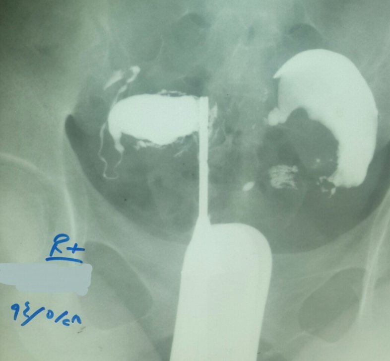
Uterus didelphys with fibroids and left tubal block
Hysterosalpingography (HSG):
- Two uterine cervices were found by initial examination.
- The left one was first cannulated, large uterine horn is demonstrated, it shows multiple filling defects suggesting submucosal fibroids.
- The left tube is blocked.
- The other cervix was then cannulated and contrast injection reveals a second uterine horn with patent tube.
- Whenever you see a unicornuate uterus, look for another cervix to rule out uterus didelphys. The other cervix is not necessarily easily seen by examination.
- If uterus didelphys is associated with obstructed longitudinal vaginal septum, only one cervical Os. may be depicted, and it may be cannulated with the endometrial configuration mimicking a unicornuate uterus.