Hepatic Hemangiomas

Case Courtesy of​

Dr Manal ElRefaei

Radiology Consultant, AlAhrar Teaching Hospital, Sharika, Egypt.

Case Images
Swipe Right and Left to see all images
Click on every item to show data
Abdomen Radiology
Hepatic Hemangiomas
  • Incidental detection of two hepatic focal lesions seen by ultrasound during evaluation of Abdominal pain, sent for further characterization by triphasic CT.
  • A: Ultrasound:

Two well-defined macrolobulated hyperechoic hepatic focal lesions

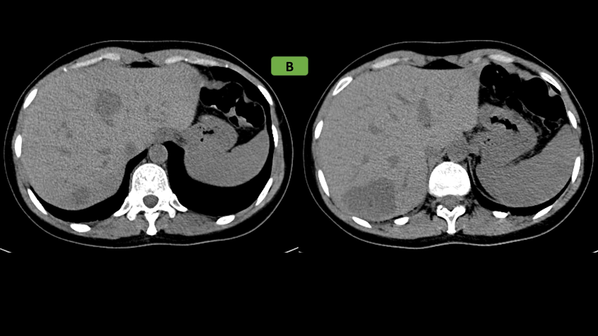
  • B: Non contrast CT:
    • Two well-circumscribed homogeneously hypoattenuating hepatic focal lesions were seen at segment IVa and VII
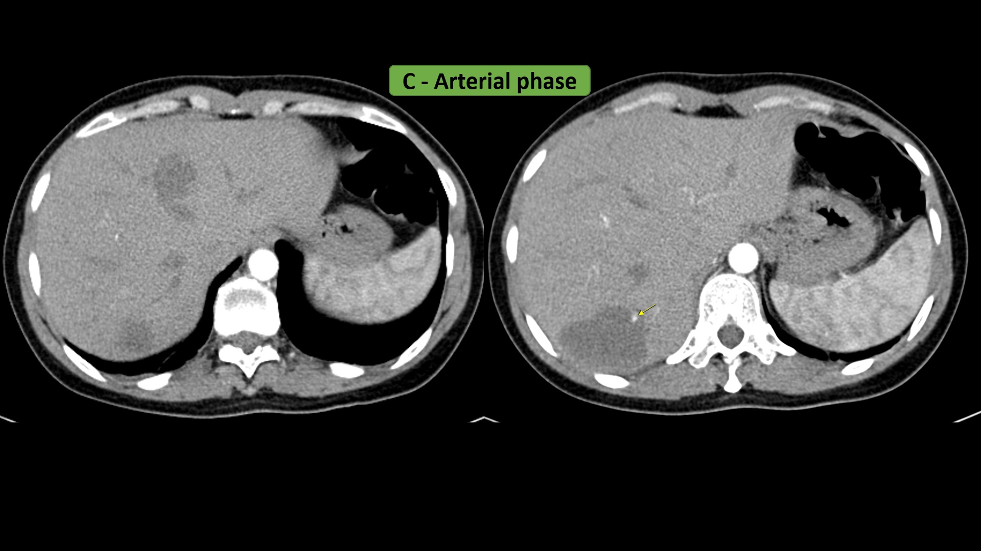
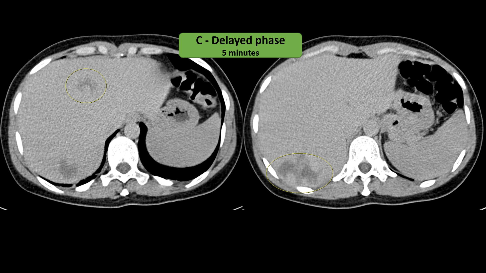
  • C: Post contrast Triphasic CT:
    • On the early arterial phase, both lesions show discontinuous, nodular, progressive peripheral enhancement with more centripetal fill-in on the portovenous phase and delayed phases.
  • It is important to differentiate hemangiomas from hepatic malignancy on different imaging modalities.
  • The discussed case shows the typical features of hemangioma on Ultrasound & CT.
  • Being familiar with the typical criteria make it easy for you to give the proper diagnosis.