
- Abdomen Imaging, Chest Imaging, Emergency Radiology, Head and Neck Imaging, Neuroradiology, Renal Imaging, Spine Imaging
- 107 (Registered)
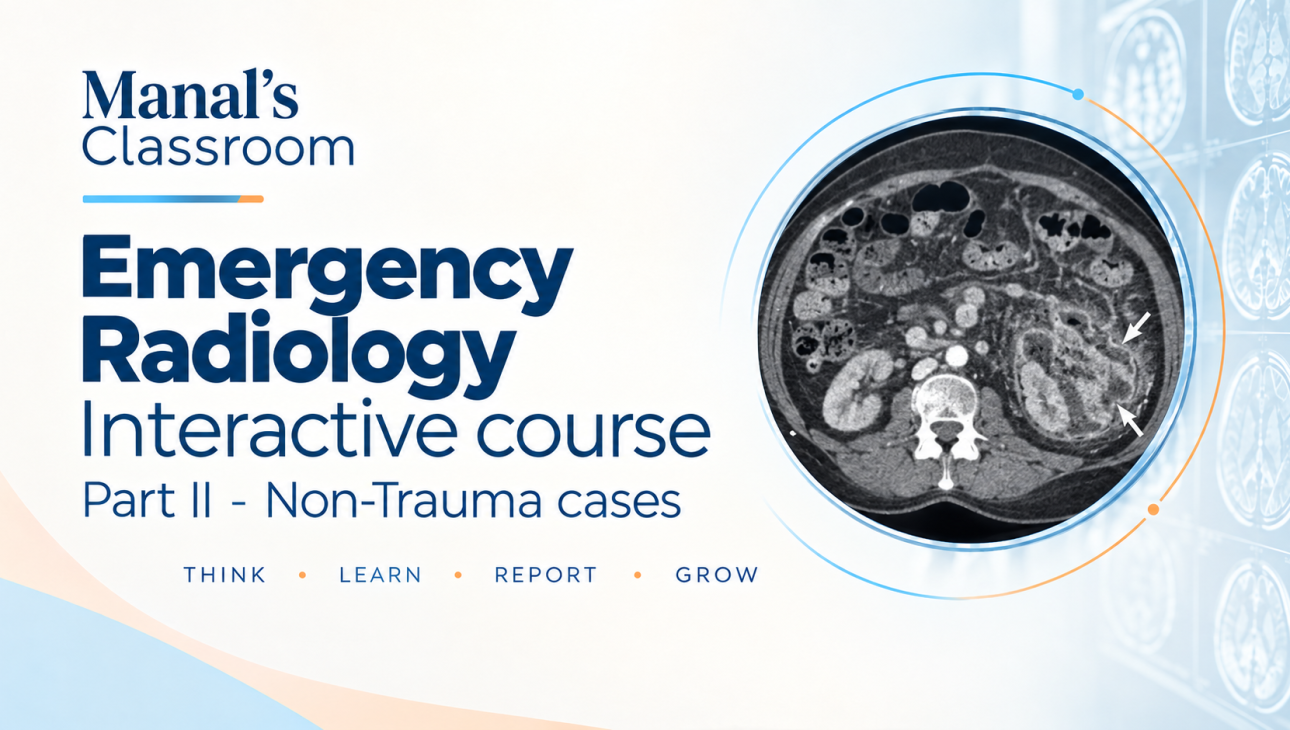
EMERGENCY RADIOLOGY INTERACTIVE COURSE – PART 2: NON-TRAUMA
Recorded Online Course
Course Instructor
Dr Manal ElRefaei, MD, EDiR, DHPE
Consultant Radiologist & Professional Medical Educator
General Organization of Teaching Hospitals and Institutes, Egypt
Target Audience
This course is designed for radiology specialists, newly appointed consultants, and early-career radiologists who wish to strengthen their approach to emergency imaging and develop greater confidence in evaluating acute non-traumatic conditions.
Course Description
The Emergency Radiology Interactive Course (ERIC) – Part 2: Non-Trauma was originally delivered as a live, case-based educational course and has been redesigned as a recorded online learning experience.
Part 2 provides a practical introduction to radiological assessment of non-traumatic emergencies, focusing on the imaging findings that radiologists need to recognize and prioritize in the acute clinical setting.
The course emphasizes the identification of life-threatening and potentially surgically significant conditions, as well as less severe findings that may be managed conservatively.
The content is designed to help radiologists develop a systematic, clinically oriented approach to non-traumatic emergency imaging, with emphasis on recognizing critical findings, understanding their clinical significance, and communicating important findings appropriately.
Throughout the course, learners are encouraged to actively test and apply their knowledge through image-based quizzes and multiple-choice questions accompanying the recorded lectures.
Course Duration
The complete curriculum is designed to be completed within 4 weeks.
The course consists of 8 recorded lectures, accompanied by image-based quiz questions and multiple-choice questions. Learners may progress through the material at their own pace while following the recommended 4-week schedule.
Learning Outcomes
By the end of this course, learners will be able to:
Select an appropriate imaging modality and technique according to the clinical emergency.
Apply a systematic approach to the radiological assessment of non-traumatic emergencies.
Recognize and prioritize life-threatening and clinically significant findings in the emergency setting.
Identify radiological findings that may require urgent surgical or medical management.
Differentiate critical findings from conditions that may be managed conservatively or electively.
Recognize important findings that should be communicated promptly to the treating clinical team.
Apply their knowledge to image-based non-traumatic emergency cases.
Course Structure
1. Pre-Test
A baseline assessment designed to explore the learner’s existing knowledge and skills before starting the course.
2. Recorded Lectures
A total of 8 recorded lectures covering key principles and commonly encountered non-traumatic emergencies in radiology.
Each lecture is accompanied by:
Image-based quiz questions
Multiple-choice questions (MCQs)
The questions are designed not only to assess knowledge but also to reinforce important concepts and introduce clinically relevant learning points.
3. Post-Test
A final assessment designed to evaluate learning and knowledge acquisition following completion of the course.
Course Completion Requirements
To successfully complete the course and receive a certificate, learners are required to:
Complete the pre-test.
View at least 80% of the recorded lectures.
Attempt at least 80% of the accompanying quiz and MCQ questions.
Complete the post-test.
Learners who fulfill all course completion requirements will be eligible to receive a course completion certificate, delivered to the email address provided during registration.
Course Curriculum
8 Recorded Lectures
Each lecture is accompanied by:
Image-based quiz questions
Multiple-choice questions
The assessments are designed to reinforce learning, test clinical application, and highlight important emergency radiology findings.
First Online Publication
8 August 2017
Curriculum
- 11 Sections
- 9 Lessons
- 4 Weeks
- Tutorial and Registration1 Item
- Pre course Radiology Quiz1 Item
- 01 Non-Trauma – Brain Imaging3 Items
- 02 Non-Trauma – Spine Imaging3 Items
- 03 Non-Trauma – H & N Imaging3 Items
- 04 Non-Trauma – Chest Imaging3 Items
- 05 Non-Trauma – Abdomen Imaging3 Items
- 06 Non-Trauma – Female Pelvis Imaging3 Items
- 07 Non-Trauma – Acute Scrotal Imaging3 Items
- 08 Non-Trauma – Pediatric Imaging3 Items
- Post course Radiology Quiz1 Item
Instructor

Related Courses
 Teacher
Dr Manal ElRefaei
 Teacher
Dr Manal ElRefaei