Pleural T-Cell Lymphoma

Case Courtesy of​

Dr Neven R. Fares

Radiology Consultant , Minia Oncology Center, Egypt.

Case Images
Swipe Right and Left to see all images
Click on every item to show data
Chest Radiology
Pleural T-Cell Lymphoma [final diagnosis by thoracoscopy and pleural tissue biopsy as well as immunophenotypic study].
  • A 30-year-old male presented by dyspnea and shortness of breath. On auscultation there was no air entry on the left, so CT chest was requested for evaluation. No history of any pleuro-pulmonary disease.

Post-contrast CT:

Contrast-enhanced Chest CT:

Coronal mediastinal window

  • Left-sided irregular circumferential soft tissue pleural thickening with nodularity involving all pleural reflections and major fissural reflection [orange arrows] with mild amount of effusion.
  • Axial mediastinal window, at the level of the heart…..Minimal pericardial effusion [white arrows].

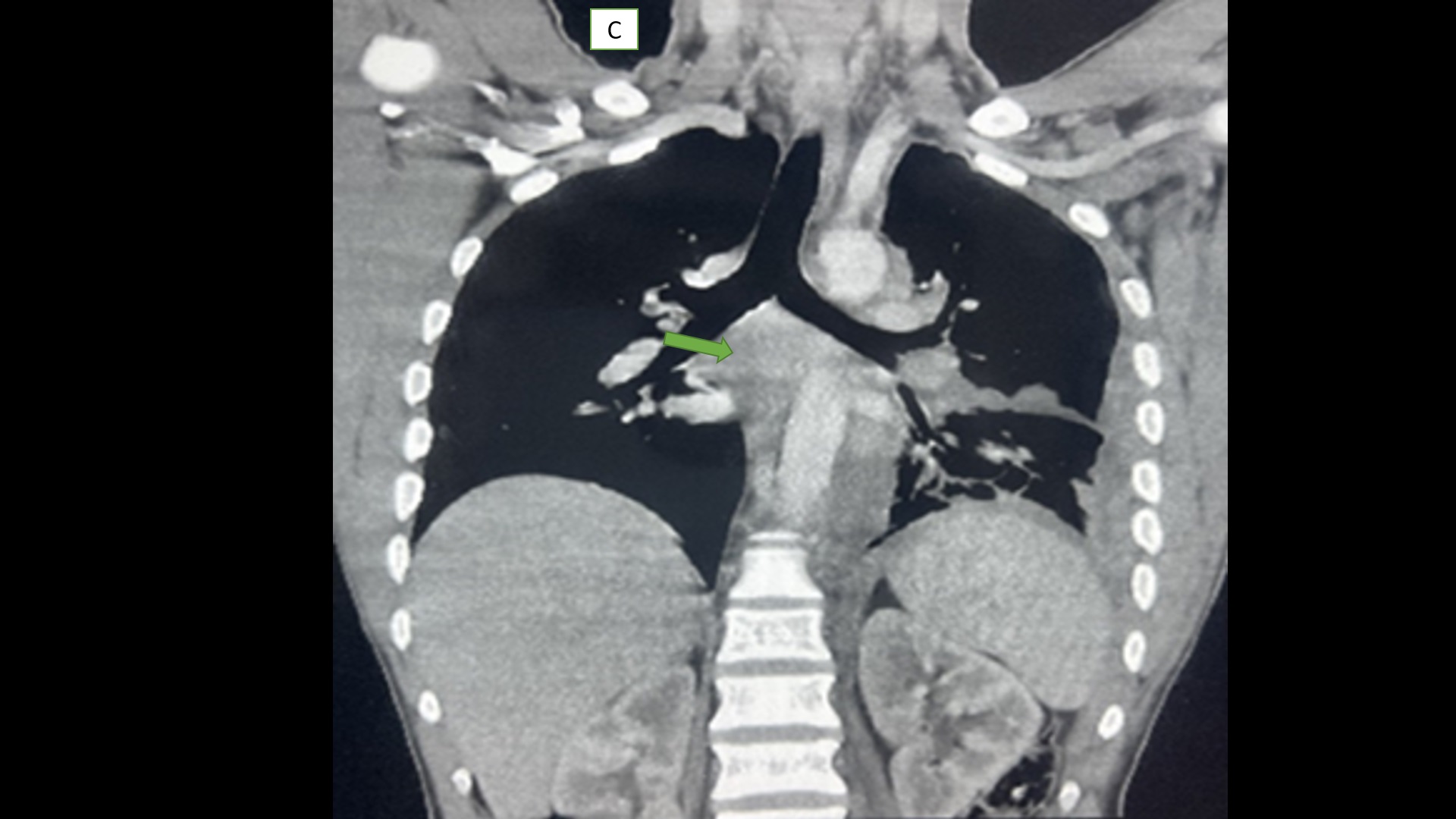
Coronal mediastinal window

  • Middle mediastinal nodal masses [green arrow].

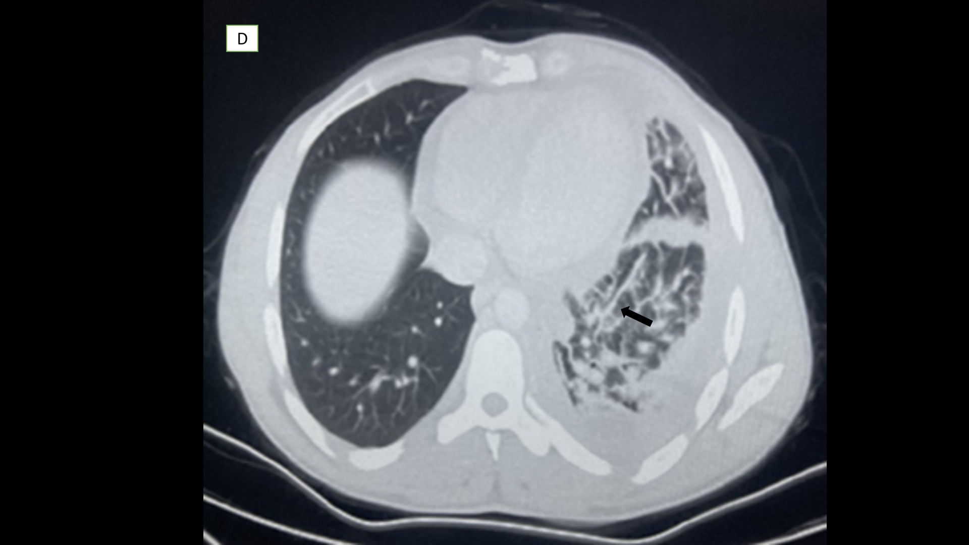
Axial pulmonary window

  • Left-sided peri-bronchial thickening, and peri-hilar interstitial thickening [black arrow].
  • T-cell primary pleural lymphoma is a very rare subtype of non-Hodgkin pleural lymphoma and could have overlapping clinical manifestations with more common conditions.
  • The mentioned findings are not specific and other conditions such as bronchogenic cancers, metastasis, infections, chronic inflammatory processes, organizing pneumonia, sarcoidosis, and lymphangitic carcinomas may have similar findings, making biopsy necessary for the diagnosis