Musculoskeletal Radiology
– Patient (01): Insufficiency fracture – Patient (02): Healing stress fracture
- Patient (01): 70-year-old female- unable to bear weight
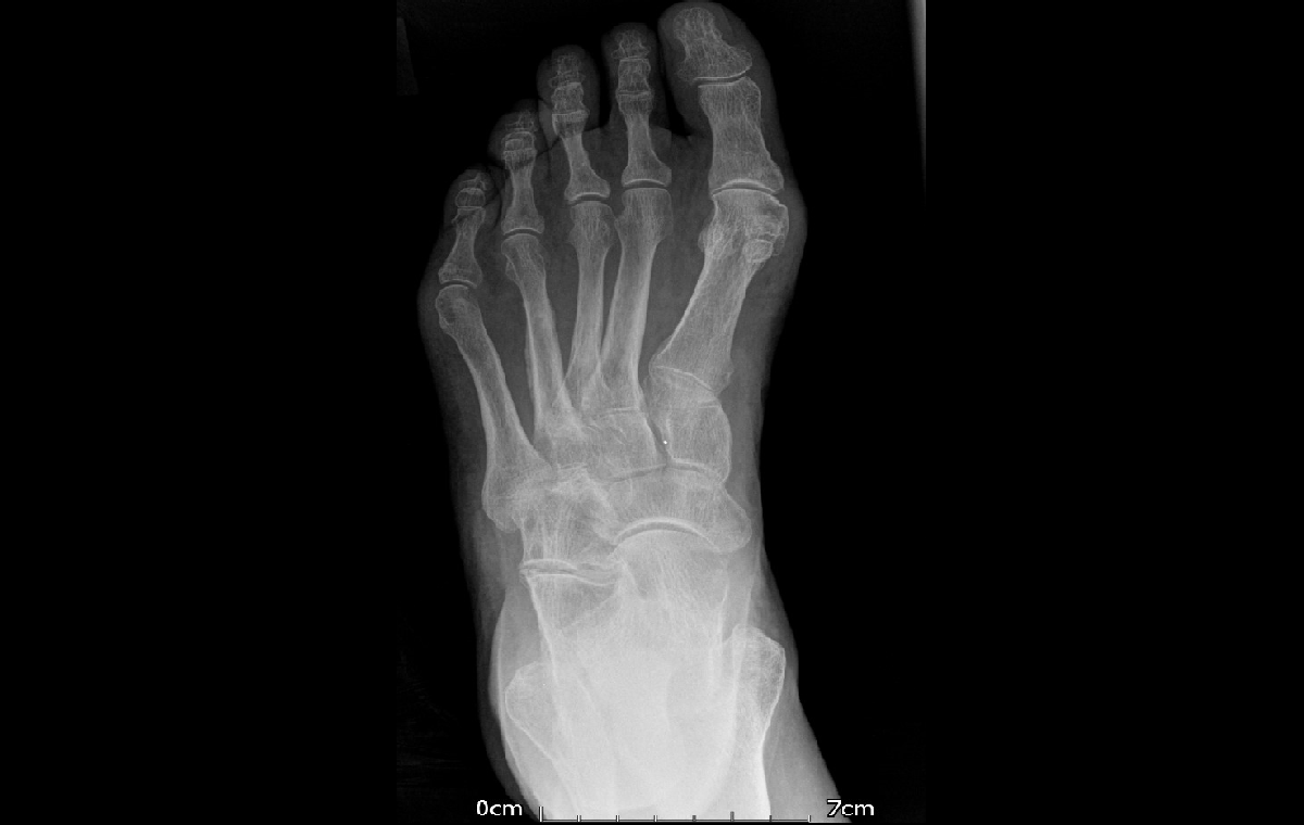
- Patient (02): 45-year-old male- left foot pain for 2 weeks – No trauma
- Patient (01):
- Osteoporosis
- A vertical sclerotic line perpendicular to the trabeculae along the posterior calcaneus.
- Patient (02):
- There is a thin periosteal reaction along the shaft of the 4th metatarsal bone consistent with a healing fracture
Patient (01):
- The calcaneus is the second commonest site of stress fracture in the foot, following the metatarsals.
- Calcaneal stress fractures typically occur as fatigue fractures in athletes involved in running/jumping or as
insufficiency fractures in the setting of RA, neurological disorders or osteoporosis. - Radiologically; occult fractures can be diagnosed by MRI seen as posterior vertically oriented fracture lines with variable bone marrow edema.
Patient (02):
- Metatarsal fractures are the most common type of stress fracture of the foot.
- Usually of the fatigue type where excessive new stresses over the normal bones result in a fracture ( Marsh fractures).
- In the acute setting, the fracture may not be visible.
- A follow-up radiograph in 10-14 days is usually recommended to check for periosteal new bone.