Neuroradiology (Emergency).
Right vertebral artery dissection & Left ICA spontaneous acute dissection (Patient is under investigation for Marfans syndrome).
- 61 years old patient with sudden onset Diplopia, mild ataxia, raised Cholesterol and Blood pressure…? evidence of posterior circulation ischemia.
- Patient had routine MRI scan that showed no evident acute infarct or hemorrhage.
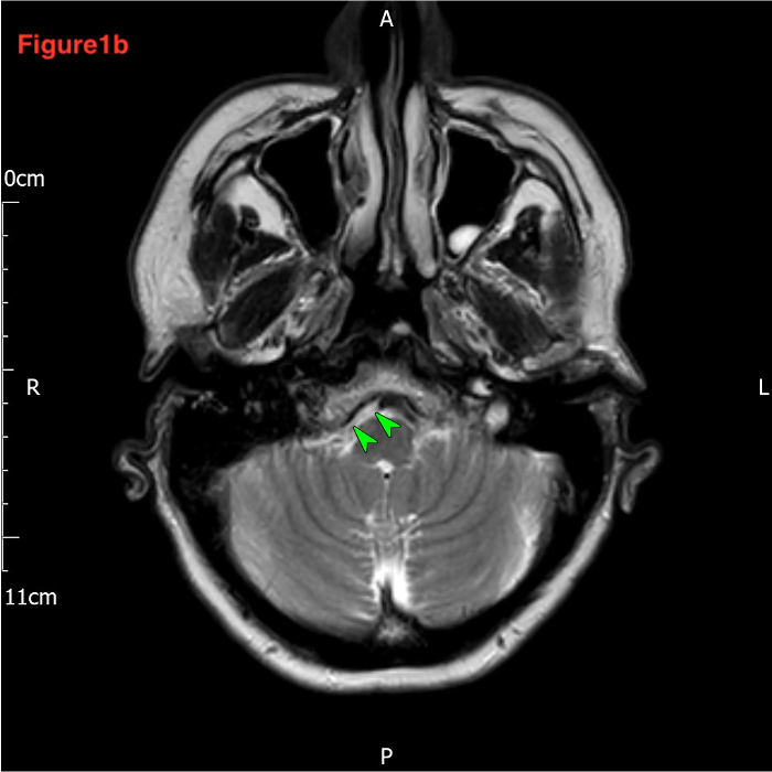
- As noted in Axial T2 (figure 1 a &b), when you compare the right vertebral and left vertebral arteries, the right one is significantly attenuated.
- Axial T2 and DWI (Figure 2 a&b) show left ICA high signal crescent seen in its wall (crescent sign).
- CT angiography was done for confirmation (Figure 3 a&b): Abnormal vessel contour of the right vertebral artery and left ICA.
- Check major cranial vessels at the Skull base (review area) even in conventional MRI.