Genitourinary/oncology radiology
- A case of operated cancer endometrium, last follow up for 4 years.
CT findings:
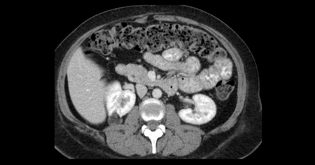
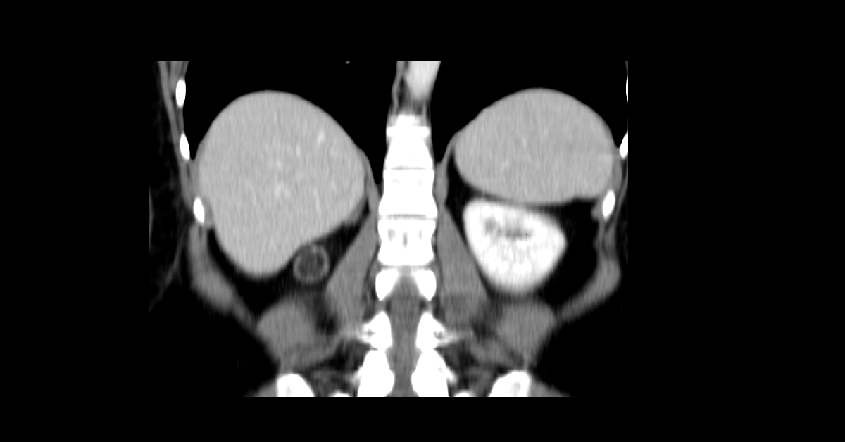
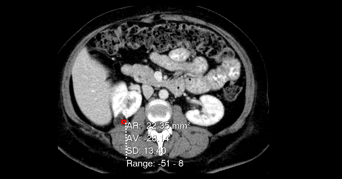
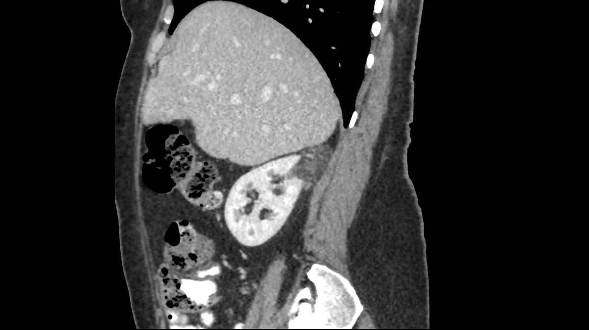
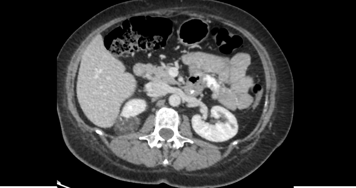
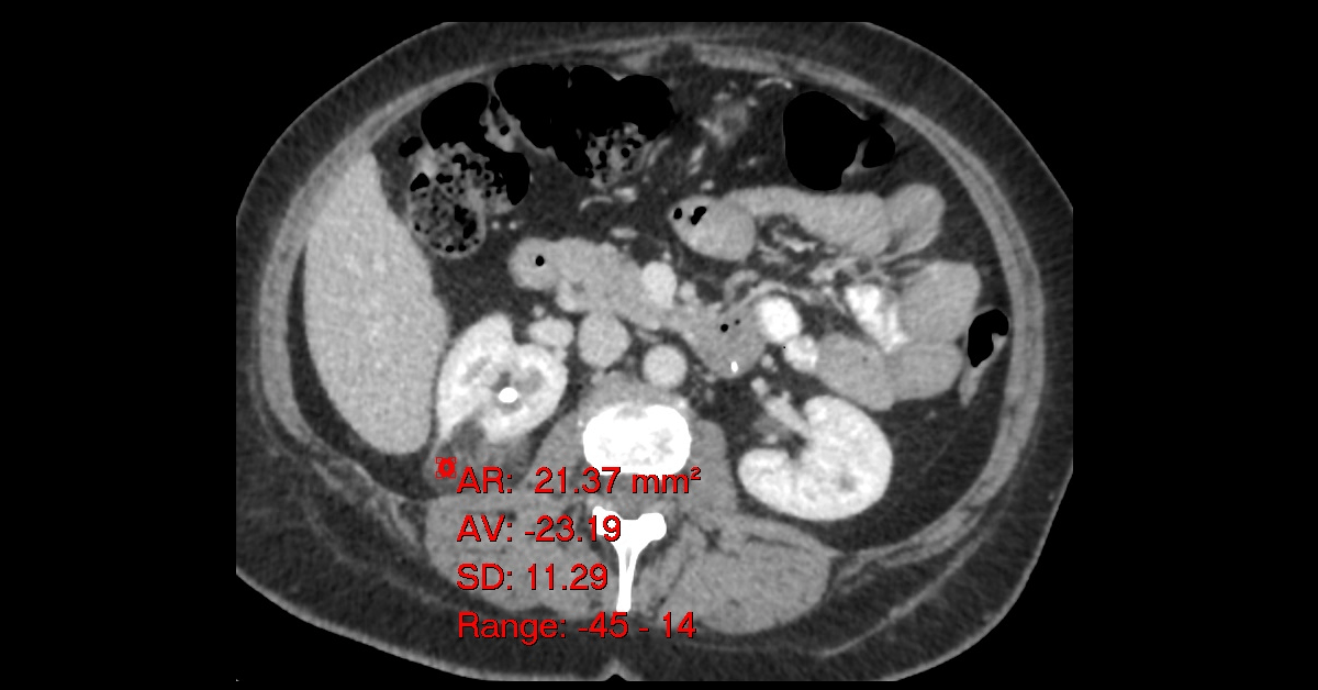
- As compared to the previous CT study done 4 years back, there is a progression in size of the previously noted well defined, faintly enhancing soft tissue mass seen exophytic from the upper pole of the right kidney posteriorly, containing gross fat (HU = -23), impressive of angiomyolipoma.
- Currently it measures 4.4 x 3.2 x 4.1 cm in TS, AP, & CC dimensions compared to 2.3 x 2.6 x 2.3 cm previously.
- This increase in size warrants surgical consultation.
- Demonstration of fatty attenuation in a renal tumour on computed tomography (CT) studies is virtually diagnostic of angiomyolipomas.
- Although it is a benign tumour, surgical consultation should be advised in patients with angiomyolipomas > 4 cm, due to increased risk of haemorrhage.