Perforated Duodenal Ulcer

Case Courtesy of​

Dr / : Ahmed Abdelfattah Mostafa

Radiologist Registrar at National Care Hospital, Riyadh, KSA.

Case Images
Swipe Right and Left to see all images
Click on every item to show data
Gastrointestinal Radiology
Perforated Duodenal Ulcer
  • A 62 years old male presented with sudden acute abdominal pain.

CT abdomen and pelvis with Oral and IV contrast.

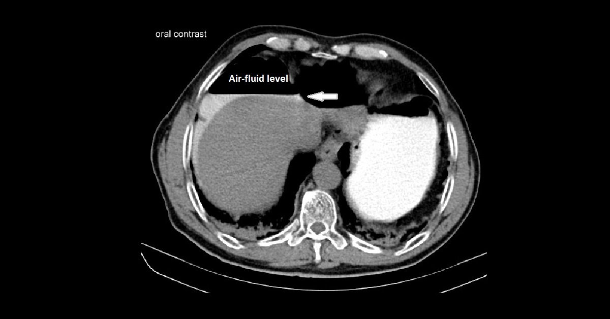
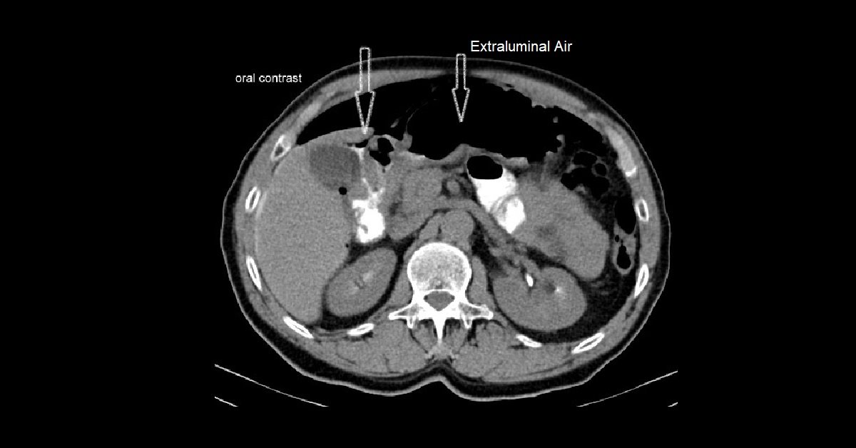
  • Marked pneumo-peritoneum with multiple intra-peritoneal discrete small hypodense pockets with air density.
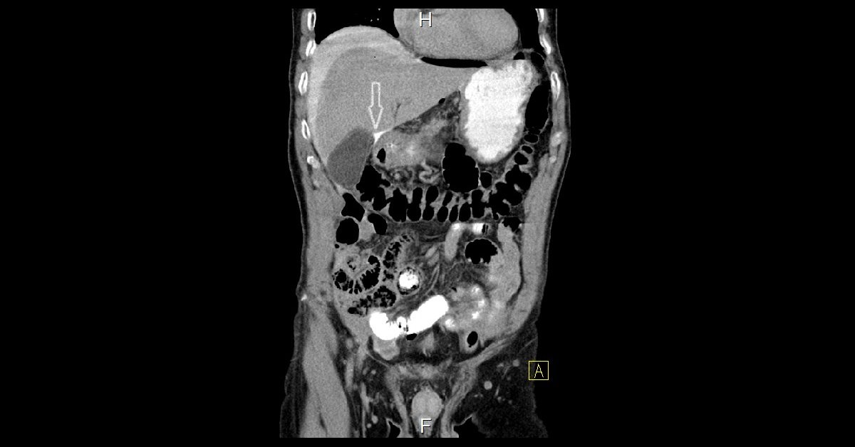
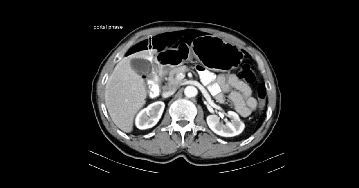
  • Ventral to the duodenal bulb and antrum are small collections of extraluminal gas and oral contrast medium  thus confirms an ulcer as the source of perforation.
  • Contrast is seen passing from a defect at these part of the duodenum on its anterior surface, medial to GB bed with contrast leakage around liver and air fluid level is detected

-Duodenal ulcer when located at the anterior wall it is perforating; while on the posterior wall it is penetrating ulcer.

– Signs of penetration and perforation

  • Wall thickening, luminal narrowing of duodenum.
  • Infiltration of surrounding fat or organs (pancreas).
  • Extra luminal, intra- or retroperitoneal gas ± enteric contrast medium.
  • Presence of intra- and extra peritoneal gas in upper abdomen is essentially diagnostic of perforated duodenum.