Gastrointestinal/oncology radiology
Multiple hepatic hemangiomas (some are giant hemangiomas)
- A case of multiple myeloma with altered liver function tests.
- Ultrasound revealed multiple hepatic focal lesions.
CT findings:
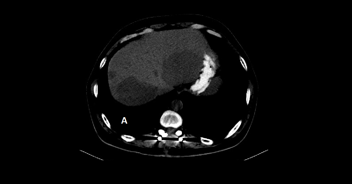
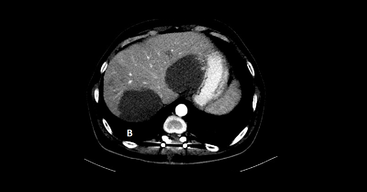
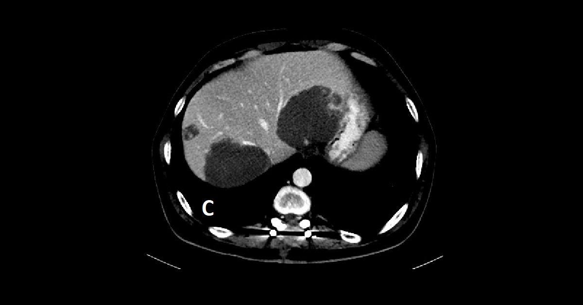
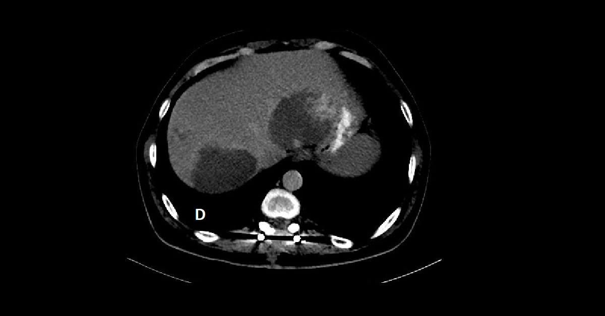
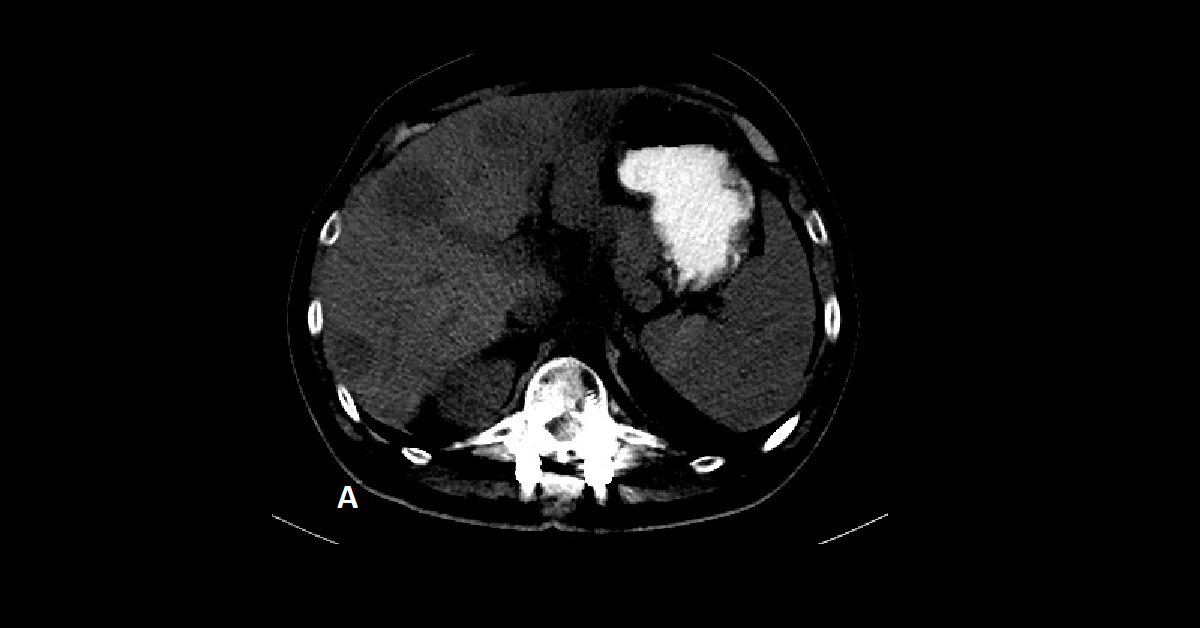
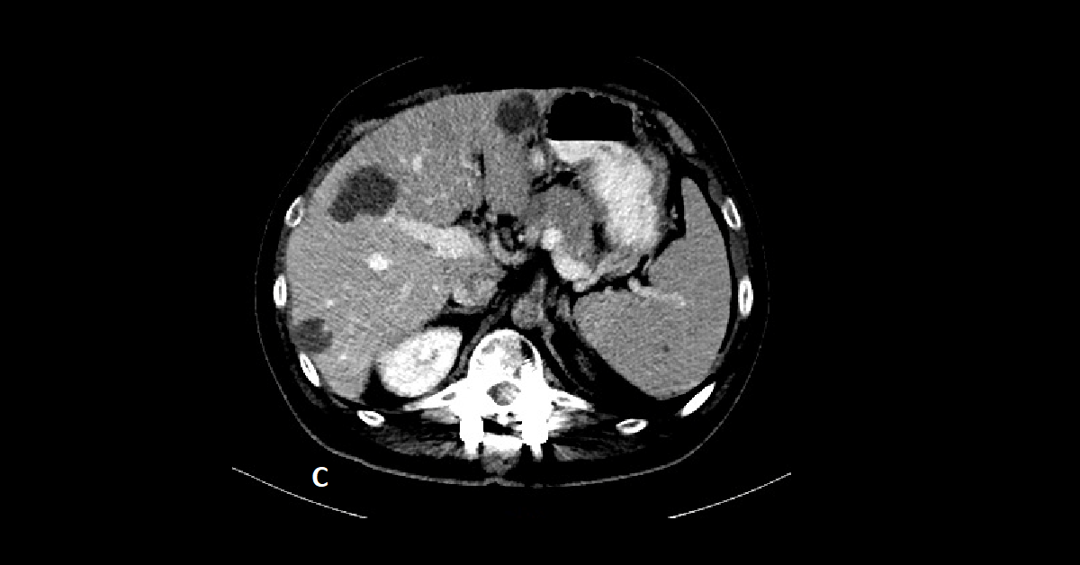
- Quadriphasic CT study (A = pre-contrast, B = Arterial phase, C = Portal phase, & D = Delayed phase) revealed multiple variable sized hypodense hepatic focal lesions scattered in both lobes, largest measures approximately 6.3 x 8.5 cm, all showing peripheral nodular enhancement in the arterial phase, with continuous centripetal enhancement in the portal and delayed phases.
- Some of the smaller lesions became more homogeneous with the liver parenchyma in the delayed phase.
- Giant hemangiomas may demonstrate similar findings to their smaller relatives, and follows aortic attenuation.
- Contrast filling may be slow and the central portions may never demonstrate complete contrast fill in.
- In such case when the diagnosis is certain, no need for further MRI assessment.
- More delayed images may be helpful.