Mal-positioned Tubes In Pediatric
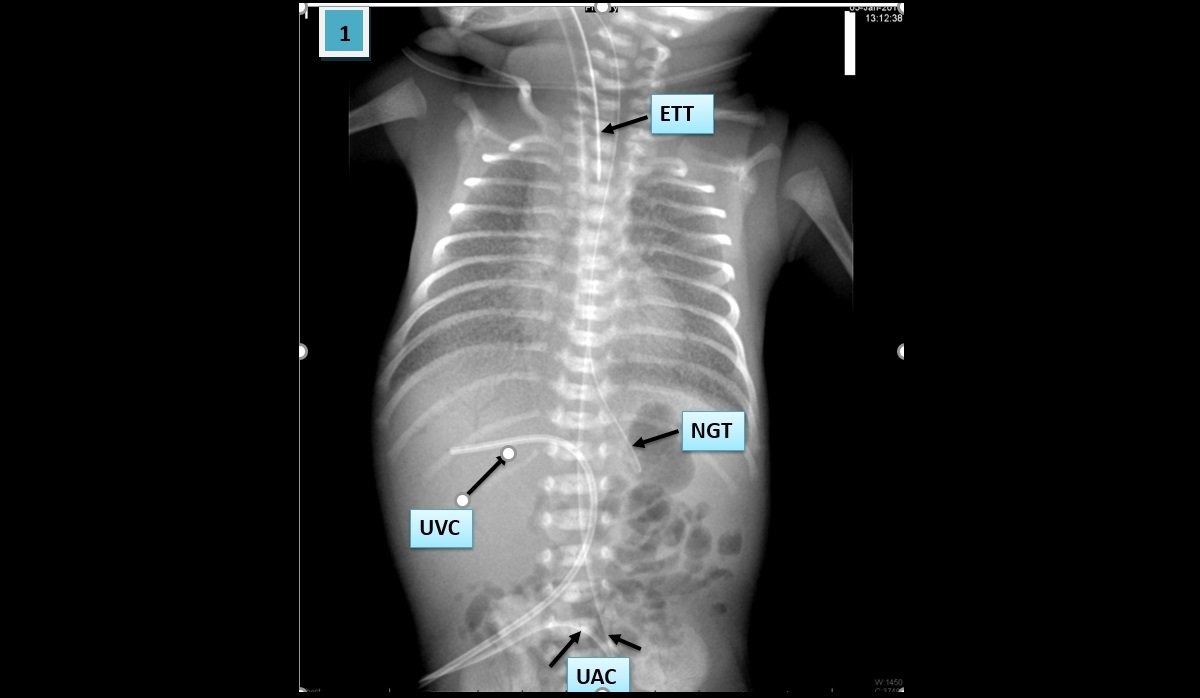
- Patient (01): Mal-positioned umbilical venous catheter (UVC) being intrahepatic into the portal venous system.
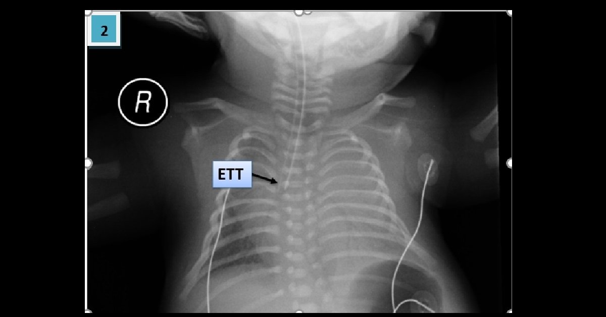
- Patient (02): Mal-positioned endotracheal tube (ETT) in the right main bronchus with left lung collapse.
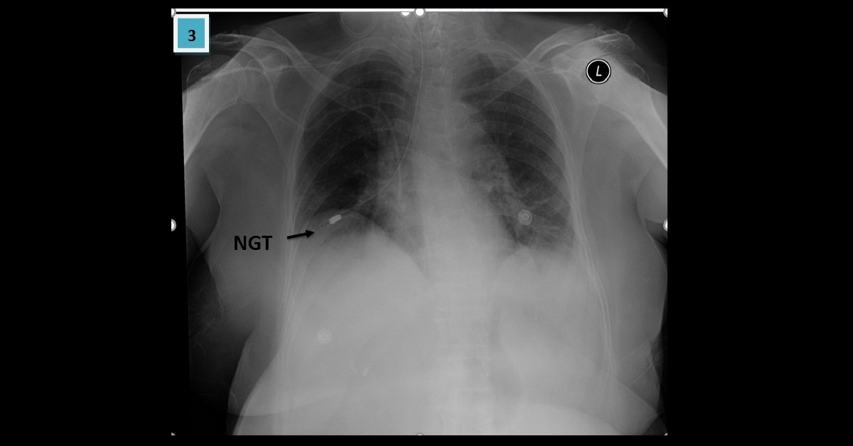
- Patient (03): Mal-positioned nasogastric tube (NGT) in the right lung.
- It is mandatory to know the imaging of lines and tubes because malposition may cause life-threatening complication for the patient.
- The tip of UVC should be positioned in the IVC above the level of the diaphragm at D8-D9 levels. Malposition in the portal circulation like our case may cause portal thrombosis.
- The umbilical arterial catheter has two types:
- High positioned: the tip should be from D6-D9 levels
- Low position (less recommended): tip level L3-L5 levels
- The NGT tip should be sub-diaphragmatic position in the stomach – identified on a plain chest radiograph as overlying the gastric bubble.
- The endotracheal tube desired position is 5 ± 2cm from the carina, but markedly varies with neck position.