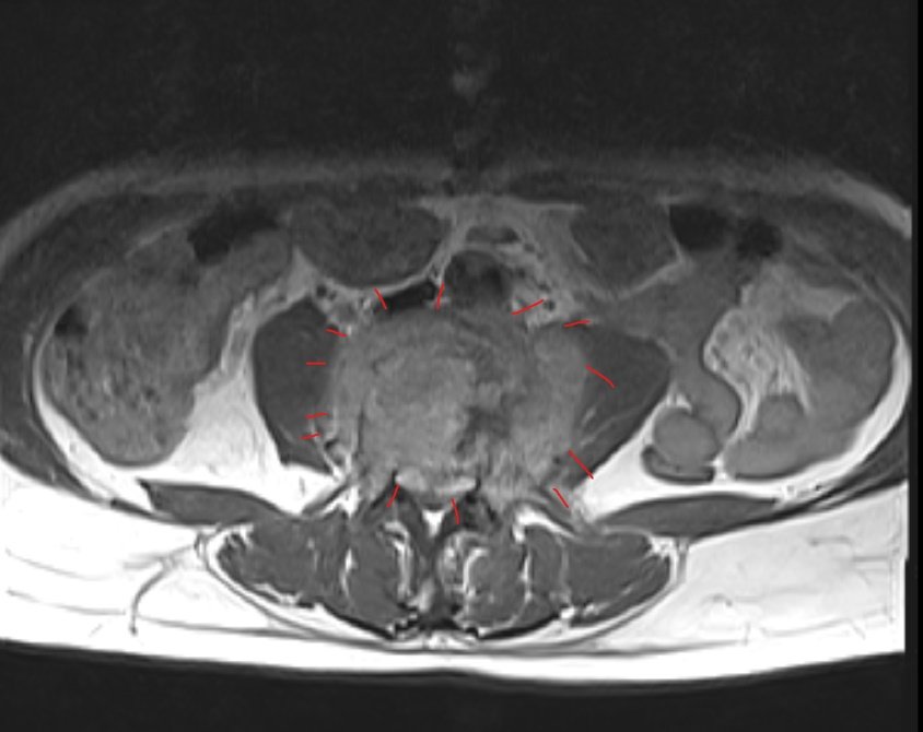
50 years old male patient was referred for CTU to assess for renal stones, but after reviewing the CTU images, dedicated history was taken: low back pain, left sciatica, intermittent fever for a month. Further evaluation by postcontrast MRI was done.
Extensive destruction and fragmentation of the L4 vertebral body [sagittal reformat].
L3-4 opposing vertebral endplates are indistinct and irregular [sagittal reformat].
Paraspinal and epidural soft tissue masses represent paravertebral and epidural phlegmons [axial images].
– Postcontrast MRI: –
Sagittal T2: shows destruction of the L3-4 endplates and L4 vertebral body. The L3 and L4 vertebral bodies show increased T2 signal, indicating oedema [orange arrows] {note green arrows of normal end plates}.
Axial T1 postcontrast: The paravertebral and epidural phlegmonous soft tissue mass enhances following IV contrast highlighting the neural compromise.