Aortic Dissection

Case Courtesy of​

Dr / Ahmed Abdelfattah Mostafa

Registrar Radiologist at National Care Hospital, Riyadh, KSA.

Case Images
Swipe Right and Left to see all images
Click on every item to show data
Vascular/Abdomen Radiology
Abdominal Aortic Dissection
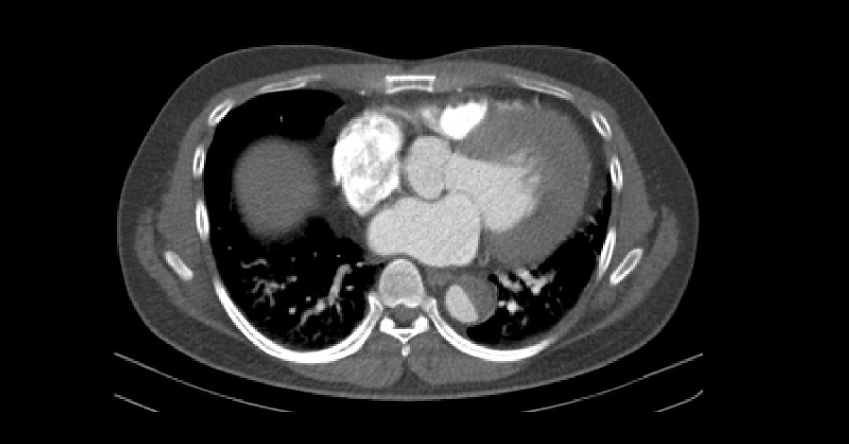
  • 49 yrs old male patient came to ER complaining of acute chest pain.
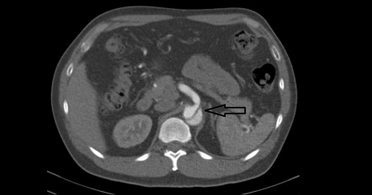
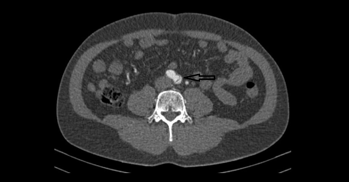
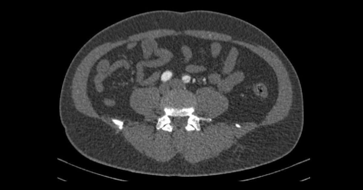
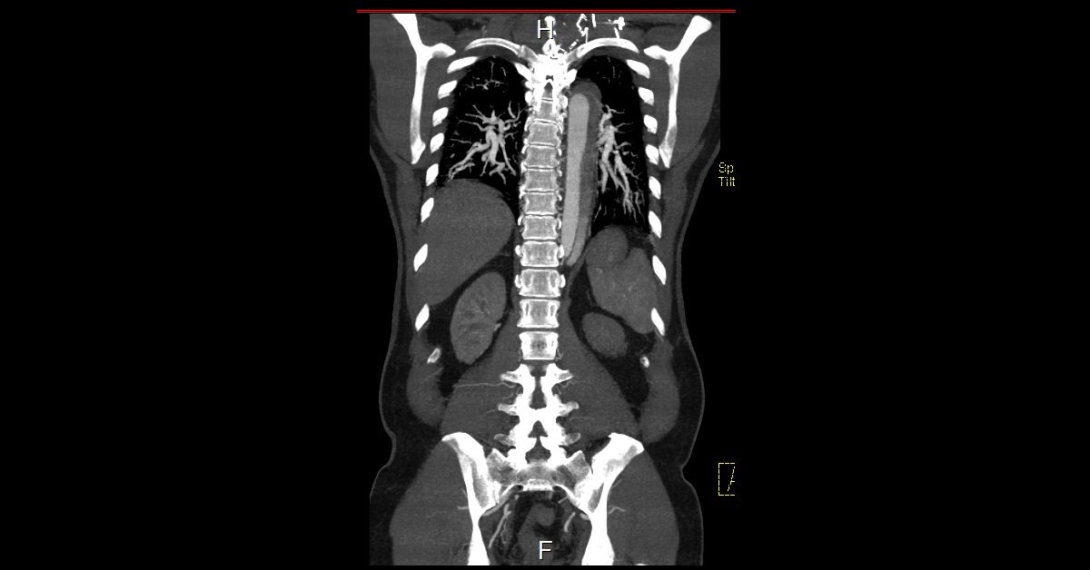
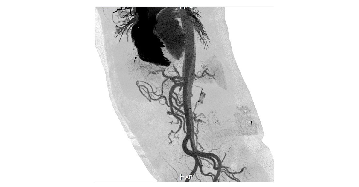
  • Evidence of intimal dissecting flap seen along the course of descending aorta extending from aortic arch just after the origin of the left subclavian artery (Stanford B) and extending down to the left common iliac artery.
  • The flap is seen abutting the origin of celiac, superior mesenteric and both renal arteries; however, no intra-arterial extension is seen showing full contrast saturation.
  • The proximal segment of the flap shows no contrast saturation…suggesting thrombosed lumen.
  • The left ventricular wall is enlarged (hypertrophied).

1-Stanford classification:

  • Type A: affects ascending aorta and arch
    • Accounts for ~60% of aortic dissections
    • Surgical management
  • Type B: Beyond brachiocephalic vessels
    • Accounts for ~40% of aortic dissections
    • Distal to the left subclavian artery

2-True lumen

    • Often compressed by the false lumen and is the smaller of the two
    • Origin of the coeliac trunk, SMA and right renal artery usually from the true lumen.

3-False lumen

    • Often larger lumen size due to higher false luminal pressures
    • Often of lower contrast density due to delayed opacification and may be thrombosed and seen as mural low density only (more common in chronic dissections)
    • Origin of the left renal artery usually from false lumen
    • Surrounds true lumen in Stanford type A.