Allergic Bronchopulmonary Aspergillosis
- In-patient admitted due to SOB with desaturation and acute exacerbation of COPD.
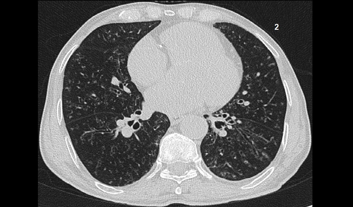
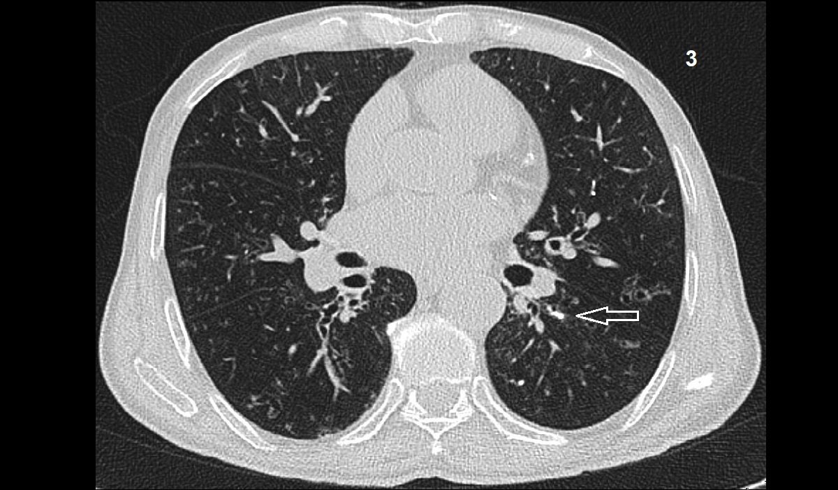
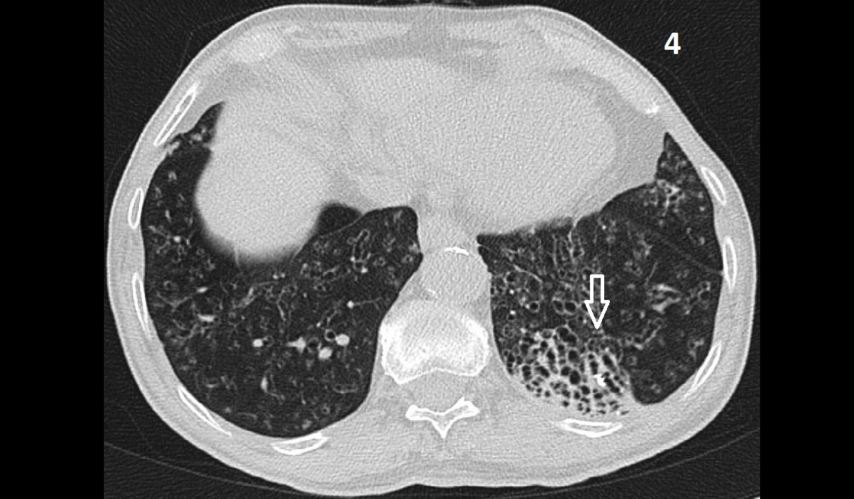
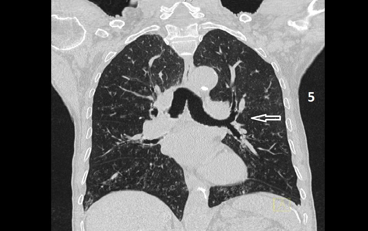
- Both lungs show diffuse mild to moderate bronchiectatic changes (Images 1,2 & 5), associated with branching calcified mucus plugs scattered in both lungs showing finger-in-glove pattern (Images 3 & 4).
- Innumerable nodular densities are seen scattered in the periphery of both lungs.
- Area of honeycombing involving the posterior basal segment of left lower lung lobe (Image 4).
- The immune mediated process is usually evident in the lung lower zones.
- The finger-in glove sign on background of bronchiectasis is highly suggestive of Allergic Bronchopulmonary Aspergillosis.