Adrenal Myelolipoma

Case Courtesy of​

Dr Sarah Ali

Radiodiagnosis Specialist - AlAhrar Teaching Hospital

Case Images
Swipe Right and Left to see all images
Click on every item to show data
Abdomen Radiology
Adrenal Myelolipoma
  • Generalized abdominal pain.
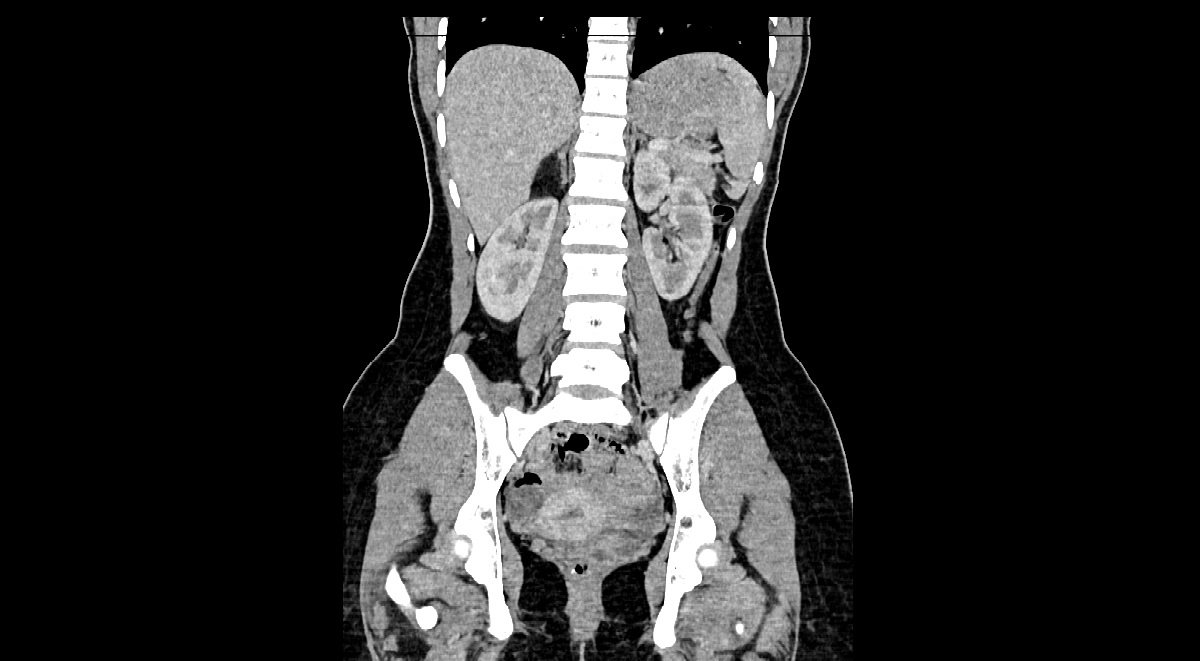
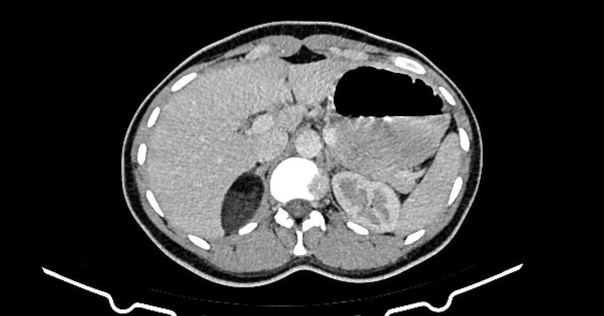
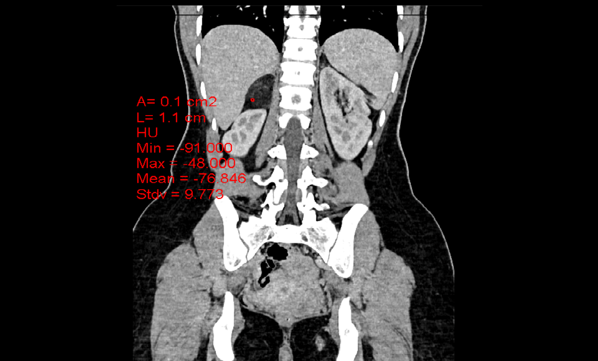
  • Axial contrast-enhanced CT images
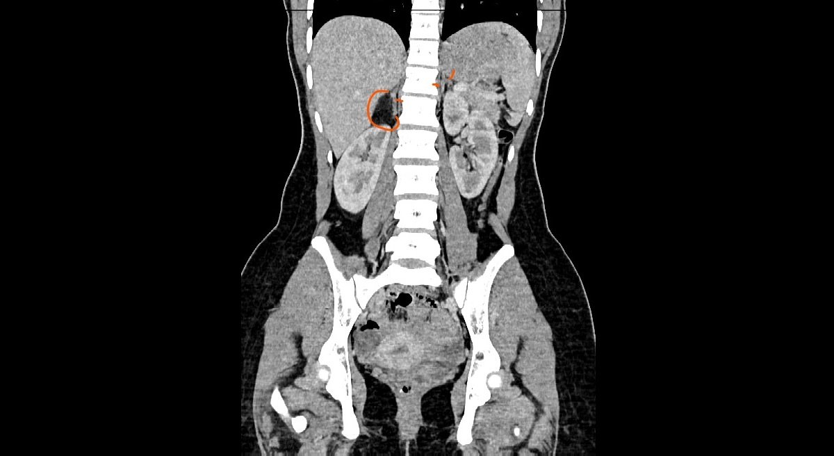
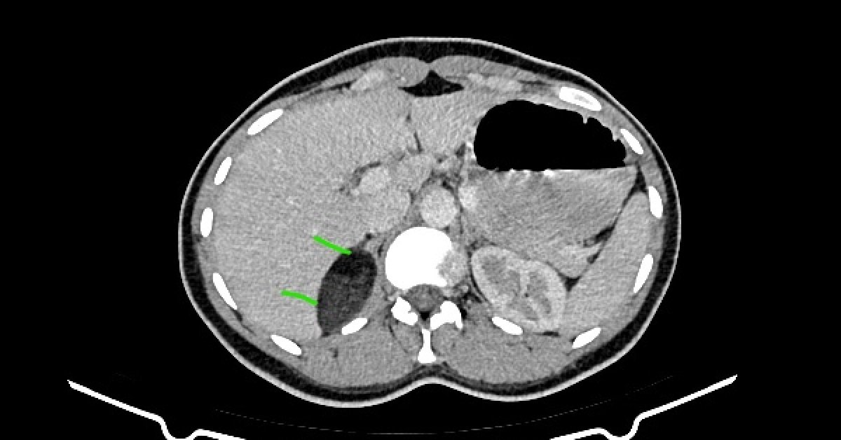
  • Right adrenal well-circumscribed mass that have mixed high attenuation {myeloid element} and low attenuation {macroscopic fat [HU= -76]} components arising from the lateral limb. It measures about 5.9 x 3 x 5 cm.
  • Orange arrows: mark both adrenal glands and the right mass that involves the lateral limb.
  • Green arrows: mark soft tissue and fat components of the lesion.
  • It is an incidental lesion, that has nothing to do with the patient complaint, don’t be misleaded.
  • The diagnosis was based on:
    • The “organ” of origin.
    • The presence of “fat” that is easily recognized helps in refining the differential diagnosis.