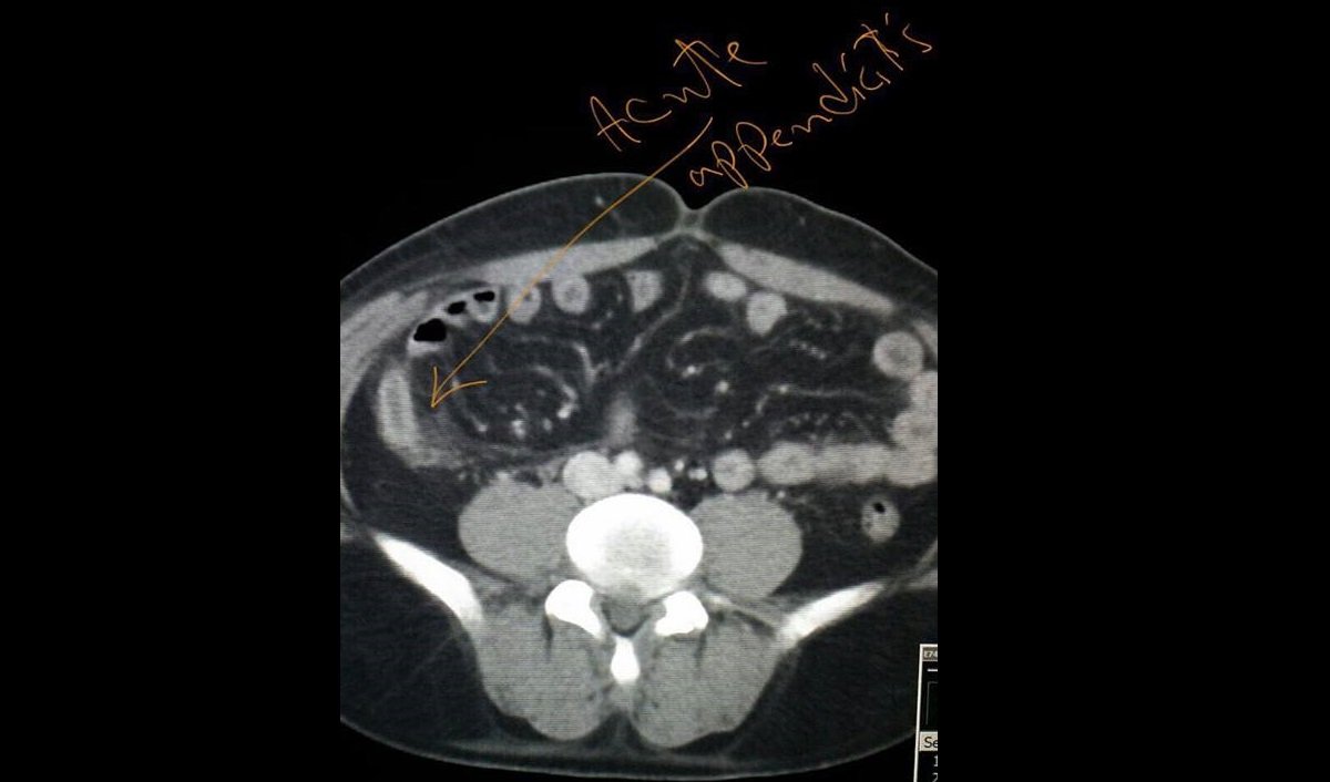
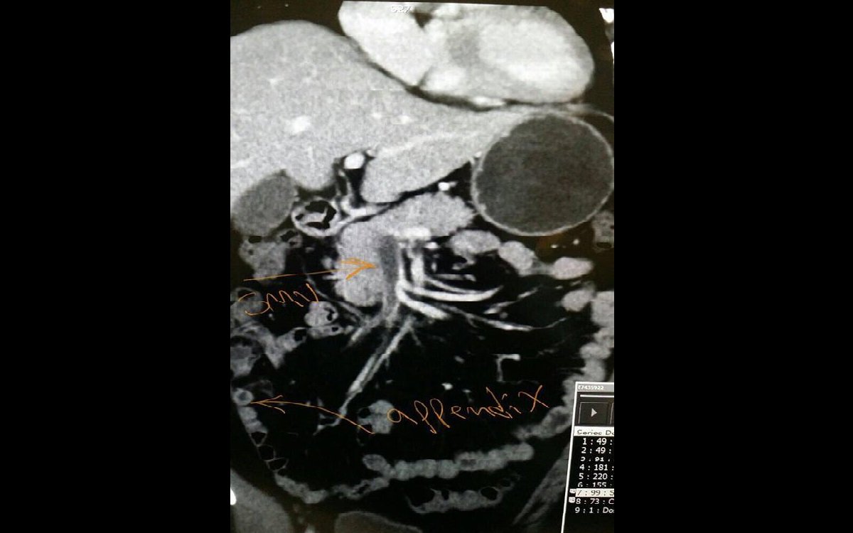
Fluid-filled thickened appendix with enhancing walls and surrounding mesenteric fat stranding, in keeping with acute appendicitis. No signs of perforation or abscess formation.
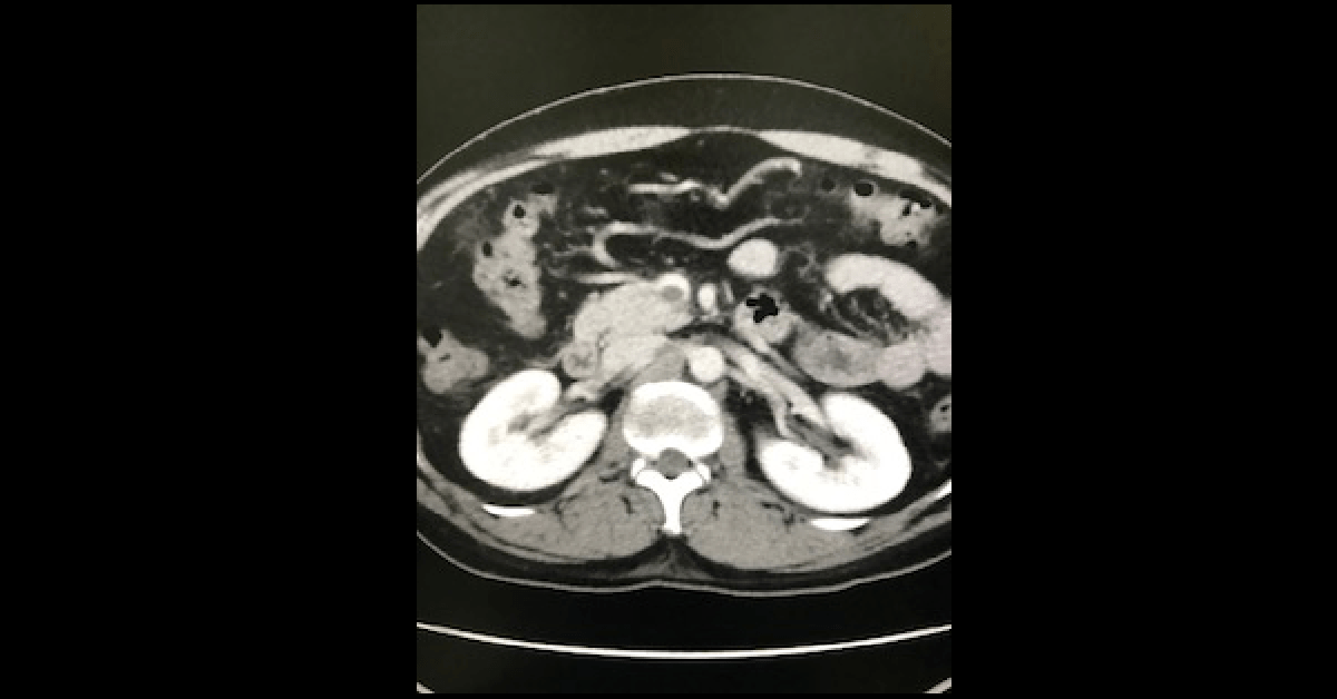
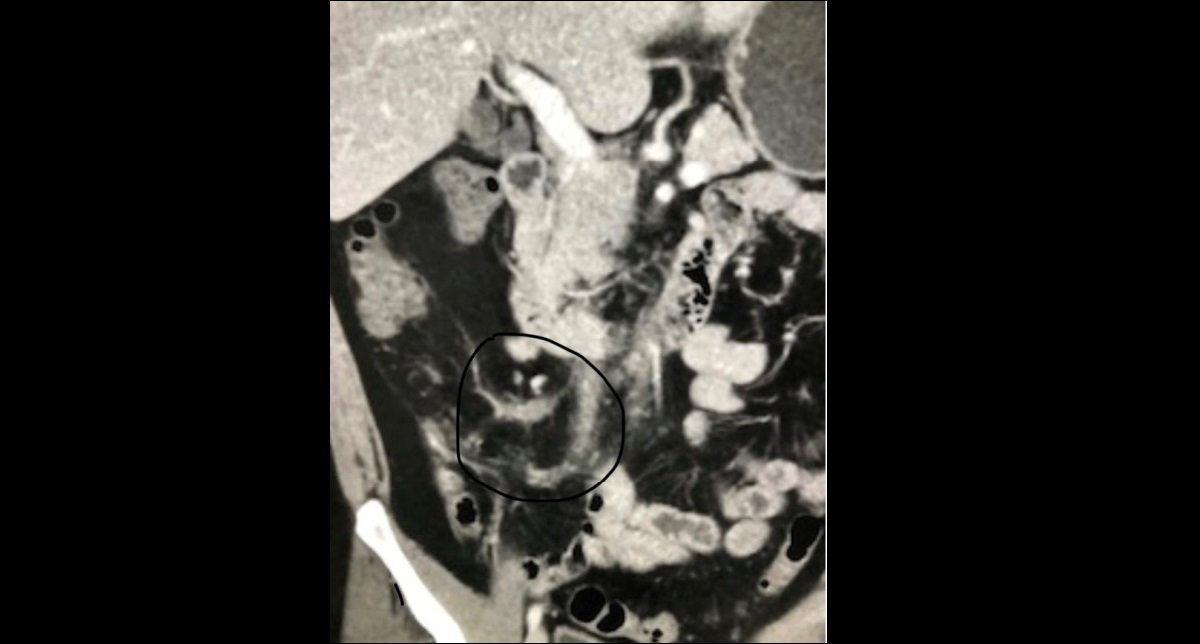
Another finding is a filling defect seen in the SMV, slightly extending into the proximal part of the portal vein and also involving few right mesenteric venous branches ( encircled in black on the image below), down to the right iliac fossa, near the site of appendicitis.
These findings suggest a thrombosis of the SMV and some of its right mesenteric branches as a complication of acute appendicitis.
A diagnosis of acute appendicitis should always prompt you to look for complications.
Always try to link your findings together: in our case, the thrombosis of the SMV is not a separate finding, but a complication of acute appendicitis, although a rare complication.