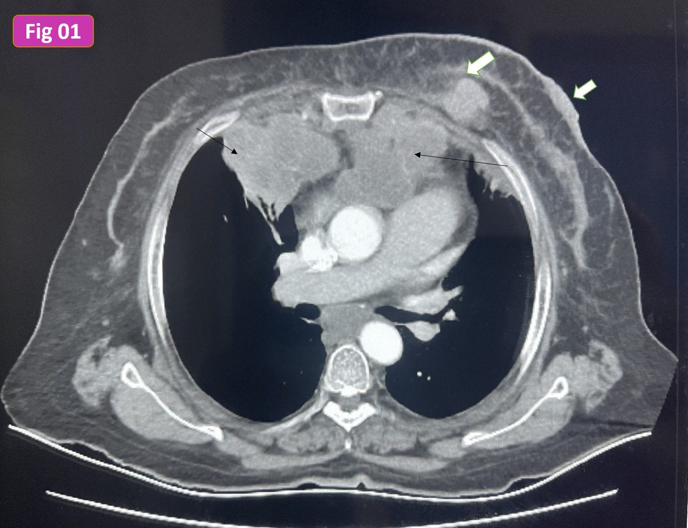
Contrast-enhanced CT chest axial cut, mediastinal window [Fig 01]: lobulated nodal masses at anterior mediastinum distributed at the region of internal mammary nodal groups bilaterally (black arrow). Incidentally detected left breast mass with mild skin thickening (white bold arrow)
Diagnostic Mammography MLO & CC views [Fig 03]: Multiple small masses scattered at both breasts with minimally increased skin thickening of the left breast (Black arrows).
Targeted Ultrasound of left breast [Fig 04]: upper inner quadrant lobulated hypoechoic mass seen para-median in location with other tiny masses seen bilaterally (white arrows)
The deeply-seated irregular mass seen at the left breast was targeted by ultrasound and a biopsy was taken.
Histopathology: undifferentiated carcinoma and Immunophenotyping revealed malignant ductal origin.
Occult breast cancers comprise a small portion of breast cancers but should be recognized.
Careful examination of the breast should be a routine part of chest CT examination and further referral to a breast imaging unit with any detected abnormality.