
Primary Pulmonary Hodgkin Lymphoma

Case Courtesy of​

Dr.Nevien R. Fares

Radiology Consultant, Minia Oncology Center, Egypt.

Case Images
Swipe Right and Left to see all images
Click on every item to show data
Chest Radiology
Primary Pulmonary Hodgkin Lymphoma

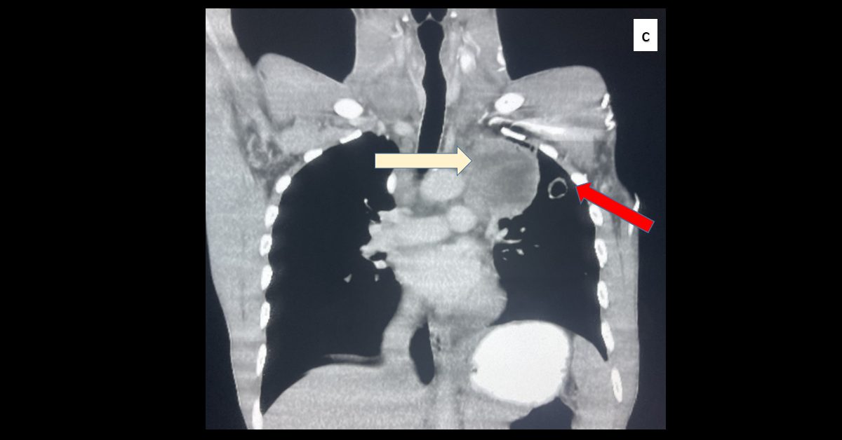
A 32-year-old male complained of a continued cough for more than 2 months and a rapidly growing neck mass for 2 weeks.

Contrast-enhanced MDCT [images A, B, C]:

  • Axial mediastinal and pulmonary windows. Coronal mediastinal window
  • Multiple variable-sized scattered cavitary lung lesions with mediastinal and hilar lymphadenopathy

– Red arrow: cavitary lung lesions

– White arrow: mediastinal LNs

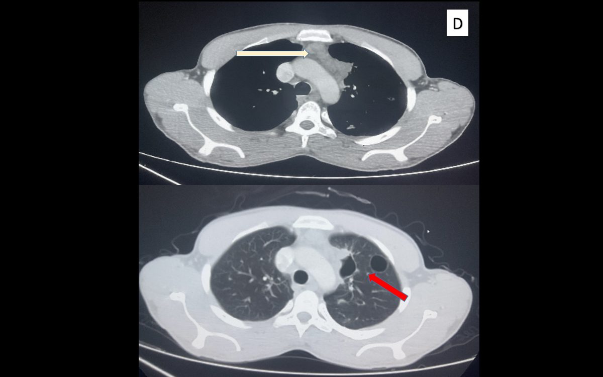
Follow-up contrast-enhanced MDCT [image D]; after 4 cycles of chemotherapy.

  • Axial mediastinal and pulmonary windows
  • Dramatic response in the form of a significant reduction in the size and extension of previously noted cavitary lung lesions and mediastinal lymphadenopathy

– Red arrow: resolved cavitary lung lesions

– White arrow: resolved mediastinal LNs

  • The rarity of pulmonary Hodgkin’s lymphoma may lead to it being not considered in the differential diagnosis of a pulmonary lesion. The frequency of lymphomas arising in the lung is estimated to be less than 1% of all lymphomas.
  • Differential Diagnosis include most importantly tuberculosis, multiple pyemic abscesses, Wegener’s granulomatosis, sarcoidosis & metastatic carcinomas, and needs to be differentiated from them
  • Diagnostic imaging plays a critical role in the initial evaluation, monitoring, and follow-up of lymphoma patients