Two well-defined macrolobulated hyperechoic hepatic focal lesions
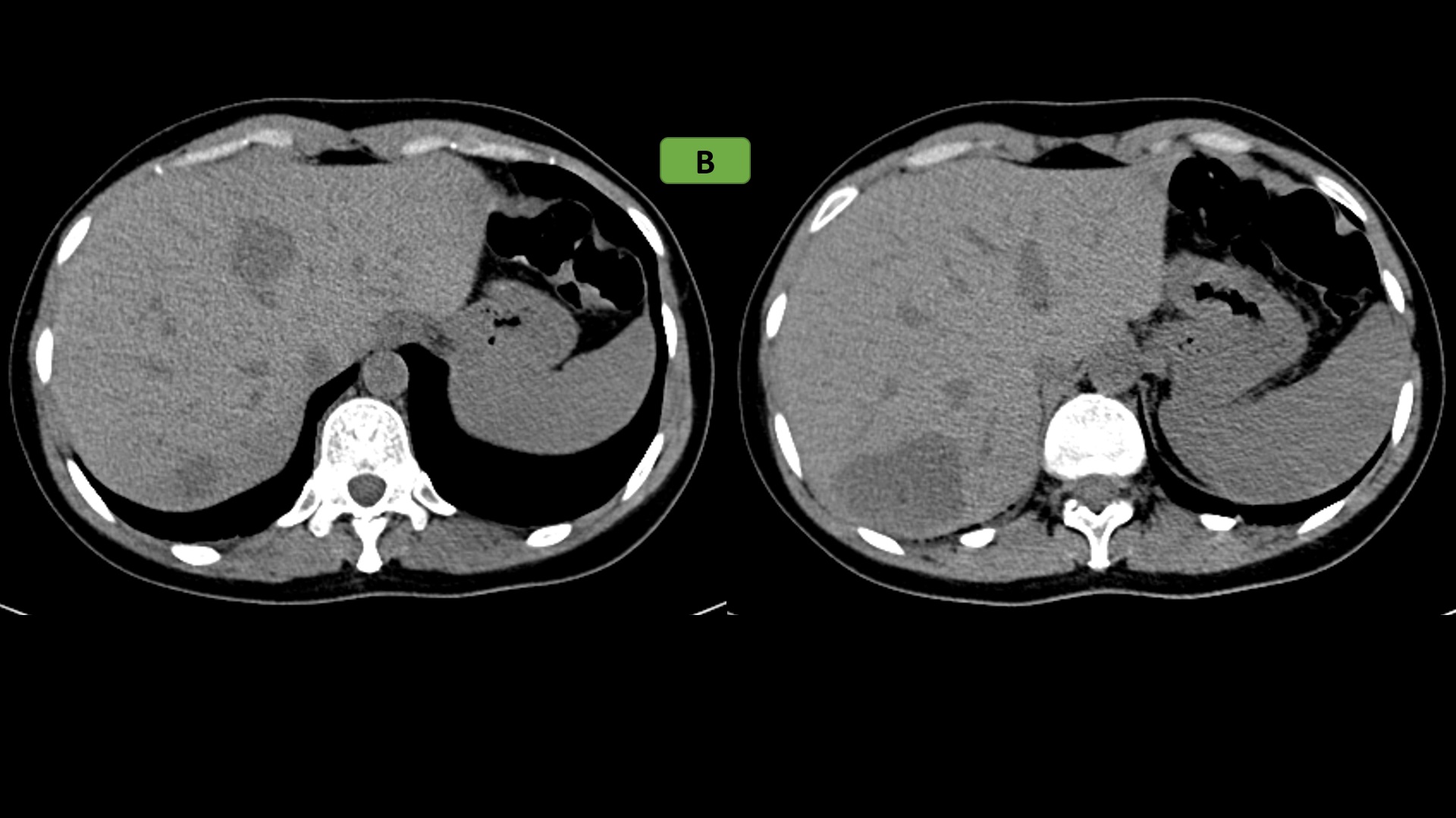
- B: Non contrast CT:
- Two well-circumscribed homogeneously hypoattenuating hepatic focal lesions were seen at segment IVa and VII
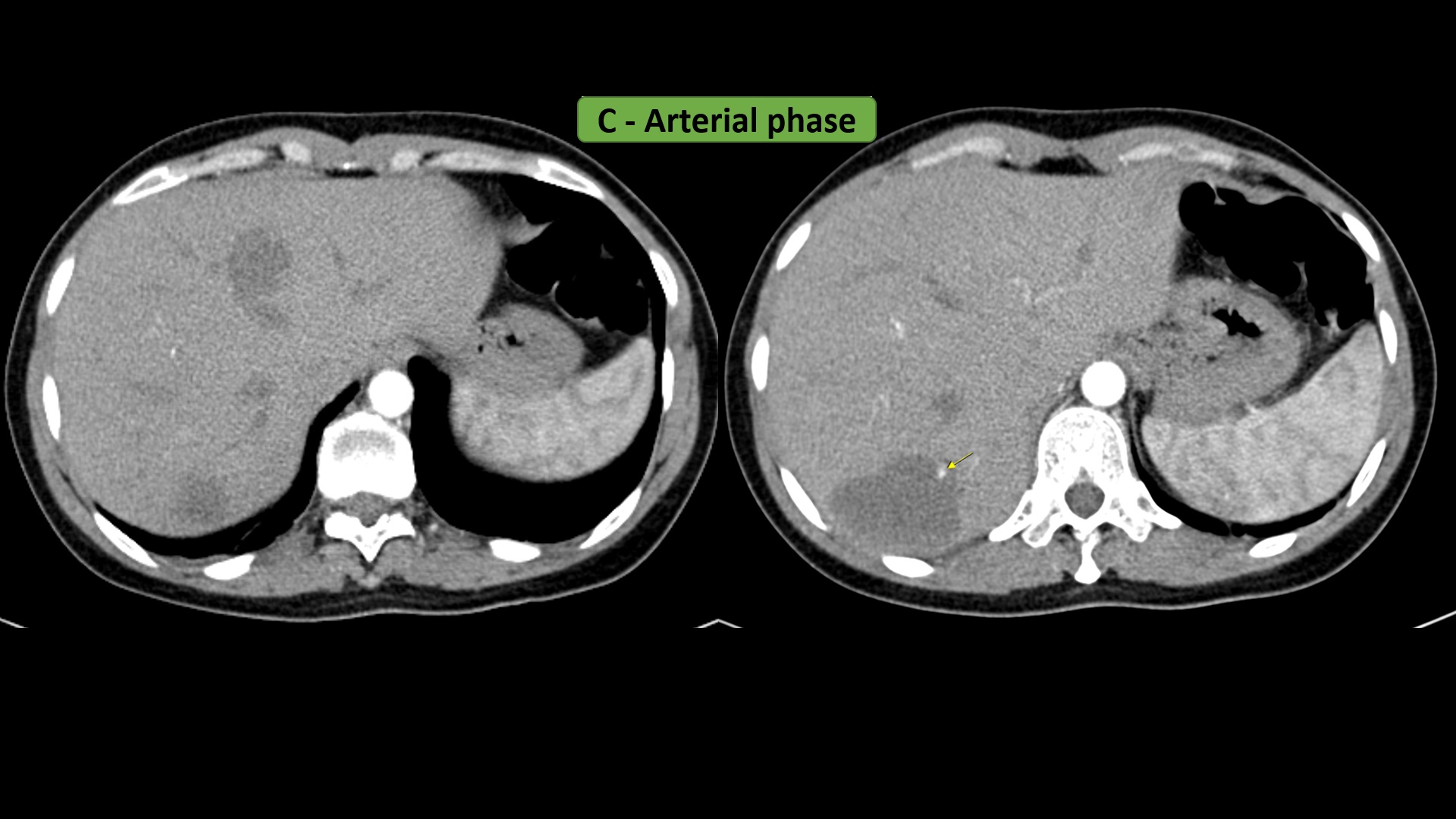
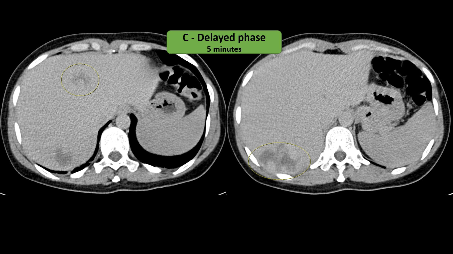
- C: Post contrast Triphasic CT:
- On the early arterial phase, both lesions show discontinuous, nodular, progressive peripheral enhancement with more centripetal fill-in on the portovenous phase and delayed phases.