Mesenteric Panniculitis

Case Courtesy of​

Dr Manal ElRefaei

Radiology Consultant - AlAhrar Teaching Hospital, Egypt

Case Images
Swipe Right and Left to see all images
Click on every item to show data
Abdomen Radiology
Mesenteric Panniculitis
  • The patient presented with abdominal pain. History of receiving treatment of Helicobacter pylori but no relief of symptoms.

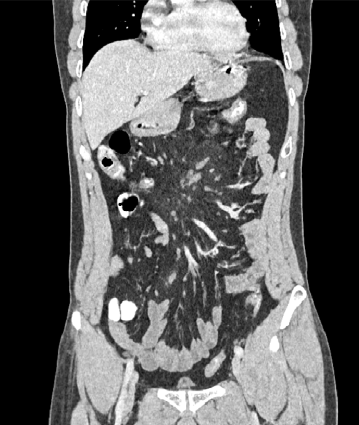
Post-contrast CT [axial and coronal cuts]:

  • A rather well defined large mesenteric mass-like lesion [red arrows] eliciting misty fat attenuation was noted. It is seen displacing the surrounding bowel loops but not displacing the surrounding mesenteric vascular structures.
  • “Tumoral pseudocapsule sign” was evident [green arrows]: it is a peripheral curvilinear band of soft-tissue attenuation that limits the heterogeneous mesenteric mass from surrounding normal mesentery.
  • Pay good attention to “mesentery” during reviewing CT abdomen.
  • Adjust the window width and length so not to miss fat-like lesions.
  • Tumoral pseudocapsule sign sensitivity is 50%.
  • The thickness of tumoral pseudocapsule is typically not greater than 3 mm.
  • When a misty mesentery is observed on CT, it is important to exclude alternative causes of a regional increase in mesenteric fat density such as oedema, haemorrhage, lymphedema, inflammation, and neoplasia before suggesting a diagnosis of MP.